Osteoarthritis of the Knee Joint
Knee osteoarthritis (osteoarthritis of the knee joint, deforming osteoarthritis of the knee joint, gonarthrosis) is a chronic, progressive disease of the knee joint based on degenerative-dystrophic changes in the joint tissues. These changes lead to the destruction of articular cartilage and underlying subchondral bone, deformation of joint surfaces, impairment of joint function, significant pain in the joint area, and a substantial reduction in the patient's quality of life.
The medical histories of patients with knee osteoarthritis are remarkably similar. Initially, minor pain appears in the knee joint area during prolonged walking. This pain subsides after a short rest. Over time, the walking distance required to trigger pain in the knee joint shortens, while the intensity of the pain increases. Patients begin to topically apply various ointments to the knee and take analgesics. After some time, they consult a physician and are diagnosed with knee osteoarthritis (gonarthrosis). In addition to pain medications, they are prescribed chondroprotectants, receive intra-articular hyaluronic acid injections, and are advised to lose weight, undergo a course of physiotherapy, and seek sanatorium-resort treatment.
Initially, this treatment provides relief. Gradually, however, the efficacy of the therapy declines, and a point comes when the pain becomes constant. When asking, "What should I do next?", patients receive the answer: "Your cartilage is wearing away, and nothing can be done about it. Walk as long as you can, and eventually, you will need joint replacement surgery".
To the logical question, "Is it possible to avoid knee arthroplasty and preserve my native joint?", the answer is affirmative: "Yes, it is possible". How can this be achieved? I will attempt to answer this question. The information presented below is based on twenty years of experience in treating knee osteoarthritis and studying the causes of its onset and progression. For many, this information may be a revelation, as it does not align with standard approaches to treating knee osteoarthritis.
The fundamental principle of medicine states: "Find the cause that triggered the disease and eliminate it; the body will then do the rest on its own".
On the internet and in widely accessible literature, numerous factors are listed among the causes contributing to the development of knee osteoarthritis: excess weight, metabolic disorders, hormonal imbalances, excessive loads on the knee, inflammation, and knee joint trauma; there are even assertions regarding a genetic nature of osteoarthritis. Our studies have shown that each of these factors occurred in specific patient groups: excess weight in 89%; metabolic disorders (excluding diabetes mellitus and gout) in 4%; hormonal imbalances in 23%; osteopenia in 67%; osteoporosis in 18%; excessive physical loads in 5%; knee joint inflammation in 2%; and knee joint trauma in 17%.
Crucially, however, 100% of the patients with knee osteoarthritis presented with an impairment of the axial load on the knee joint. Furthermore, the severity of osteoarthritic changes in the knee joint has a clear correlation with the severity of the axial load impairment on the joint. This allows us to state that the primary cause of the onset and subsequent progression of knee osteoarthritis is the impairment of the axial load on the joint. These impairments occur as a result of deformity (curvature) of the lower leg (more precisely, as a result of deformity of the tibia).
How can a tibial deformity trigger the development of knee osteoarthritis? The explanation is quite simple. We are all well-acquainted with the laws of physics—specifically Newton's third law (action equals reaction) and the laws of leverage. These laws explain everything clearly. When we walk, the force with which we press against the ground acts equally upon the articular surfaces of the knee joint. The tibia serves as a kind of lever through which this action is exerted. This lever has two arms: a long arm (the length of the tibia) and a short arm (the areas of the articular surfaces). When the lower leg is straight, the total force acting on the knee joint is evenly distributed across the articular surfaces. When the tibia is curved, the law of leverage comes into play, and the force acting on the articular surfaces is distributed unevenly. Areas appear within the knee joint that experience significantly greater loads than other areas of the joint. In the regions of the articular surfaces subjected to increased load, changes begin to develop that represent the earliest signs of knee osteoarthritis. The longer these joint areas are overloaded, the more pronounced these changes become, and the more severely knee osteoarthritis manifests.
The other factors listed above are secondary and merely contribute to either a slower or more rapid progression of changes within the knee joint. Consequently, to treat knee osteoarthritis effectively, it is necessary to normalize the axial load on the joint. This can only be accomplished surgically.
Modern principles of knee joint osteoarthritis treatment do not differ from those formulated back in the 1970s and 1980s. These principles are based on two concepts: first, conservative treatment is conducted (pharmacotherapy, physiotherapy, sanatorium-resort treatment, and therapeutic exercise), the aim of which is to prolong the time the patient can use their own joint; then, when the pain becomes debilitating, joint replacement (endoprosthetics) is performed.
It is completely clear to everyone that despite the advancement of pharmacology and the refinement of physiotherapy equipment, no conservative procedures can eliminate tibial deformity or normalize the load on the knee joint. Surgical intervention is required. However, in the initial stages of knee osteoarthritis, arthroscopic surgeries are most frequently performed. The reason is that during MRI examinations, degenerative changes in the menisci are detected in all patients with knee osteoarthritis. Therefore, patients are advised to undergo arthroscopic surgery to "clean out" the joint. However, this is a palliative surgery; it does not eliminate the root causes of osteoarthritis, and after a year to a year and a half—and in many cases even sooner—the pain in the knee joint returns. In several European countries, such as the UK, France, and Germany, performing arthroscopy for a diagnosis of knee osteoarthritis is prohibited because arthroscopy impairs metabolic processes within the joint and accelerates the progression of degenerative processes.
In our practice for treating knee osteoarthritis, we utilize a surgical technology developed by our team. This is an extra-articular surgery that eliminates the primary cause of knee osteoarthritis—the impairment of the axial load on the knee joint. Furthermore, this surgery allows for the stabilization of the ligamentous apparatus of the knee joint, which is very frequently lax ("loose") in patients with knee osteoarthritis. The normalization of the axial load on the knee joint and its stabilization create the conditions required for the normalization of metabolic processes within the knee joint and the regeneration of articular cartilage.
When necessary, as an additional factor promoting knee joint restoration, we utilize stem cells, which are introduced into the joint 3–4 weeks postoperatively. It must be noted immediately that one should not expect a rapid effect. The volume of therapeutic interventions, the duration of treatment, and its efficacy depend on the stage of the disease and the condition of the knee joint tissues. The longer the osteoarthritis has existed, the more pronounced the changes that develop in the knee joint tissues, and the more time is required for these changes to undergo reverse development.
In every patient, the changes in the knee joint have unique characteristics. Therefore, following a detailed examination of each patient, we develop an individualized treatment program. Unfortunately, however, when our examination reveals that the changes in the knee joint tissues are irreversible, we are compelled to recommend joint replacement surgery to the patient.
Over the past 15 years, our surgical technology for treating knee osteoarthritis has been successfully used in more than 170 patients. To the best of our knowledge, none of them have required knee arthroplasty.
Clinical cases
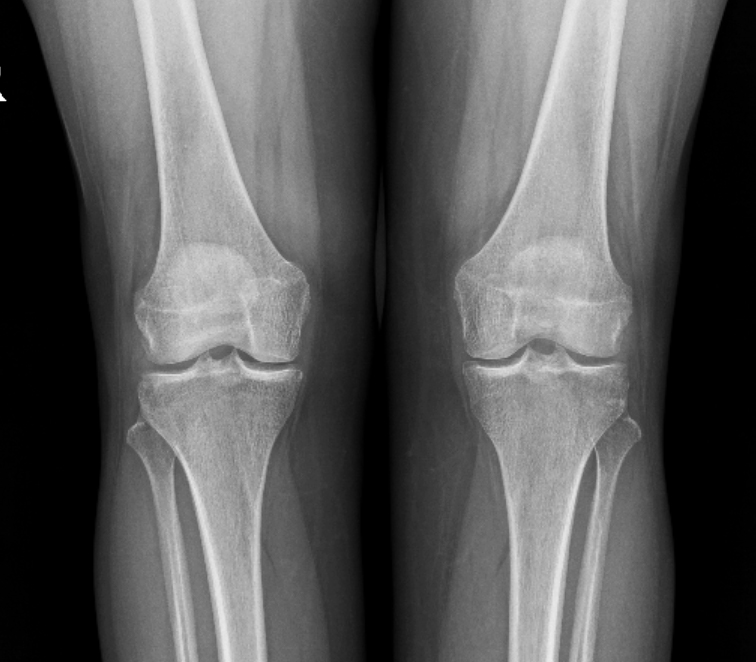
The man turned with complaints of severe pain in the left knee joint. Pain in the joint occurred immediately upon starting to walk, which forced the patient to use crutches while walking. The patient consulted at various medical institutions, all of which recommended that he replace his left knee joint with a prosthesis.
Before treatment . The radiograph (1) shows a pronounced varus deformity of the tibia. On MRI images (2) there is a section of the articular surface with a complete absence of articular cartilage (circled by a dotted line); articular fluid is detected in place of the cartilage. Normally, articular cartilage appears as a wide gray stripe (indicated by arrows).
We carried out restorative surgical treatment using autologous mesenchymal stem cells.
One year after surgical treatment. On the radiograph (3) the axis of the tibia has been restored. On MRI images (4) new cartilage up to 3 mm thick has formed in place of the previously missing articular cartilage (circled by a dotted line).
Two years later After completion of treatment, according to the patient, he leads a normal life. Walks independently without using crutches or a cane. Pain in the left knee joint does not bother him, although, as the patient himself notes, “if necessary spend the whole day on your feet, doing housework and gardening “, then in the evening a slight aching pain appears in the joint, which quickly goes away on its own after you sit in a chair or chair for 10-15 minutes.”

A 64-year-old male patient, Volodymyr, presented to our clinic presenting with osteoarthritis of the left knee joint. The initial manifestations of osteoarthritis appeared 3 years ago. One year ago, he consulted a local orthopedic trauma specialist who informed him that total knee arthroplasty was indicated and referred him to a specialized department. Volodymyr declined the surgical procedure and sought consultation from other specialists. He underwent conservative treatment, which yielded no clinical improvement. Several specialists informed him that his osteoarthritis was associated with laxity ("sagging") of the medial collateral ligament of the knee joint and displacement of the medial meniscus. This pathology causes the articular surfaces of the bones to rub against one another. They noted that reversing these intra-articular changes is impossible, which is why joint replacement is necessary. Furthermore, Volodymyr was told that at his age, it is futile to expect any meaningful outcome from osteoarthritis treatment.
Our examinations revealed that the cause of the pathological changes in the knee joint was an impairment of the joint alignment and load distribution, which was induced by a significant deviation of the force acting on the tibia away from the mechanical load axis.
Volodymyr underwent surgical treatment, during which correction of the knee joint alignment/load distribution and core decompression (tunneling) of the femur and tibia were performed. This surgical intervention established the necessary conditions for the maximum possible regeneration of the joint.

A 48-year-old female patient, Viktoriia, presented to our clinic presenting with osteoarthritis of the right knee joint. The diagnosis of knee osteoarthritis was established approximately 5 years ago. She had previously undergone conservative management, including analgesics, chondroprotectants, platelet-rich plasma (PRP) therapy, and intra-articular injections of hyaluronic acid. While the pain subsided temporarily, it invariably recurred. Consequently, Viktoriia was informed that she would most likely require total joint replacement surgery in the near future.
Upon objective clinical and radiographic examination, the patient was found to have a varus deformity of the right tibia, which caused the mechanical force acting on the bone to significantly deviate laterally, resulting in a substantial overload of the medial compartments of the right knee joint. An MRI examination revealed bone marrow edema (so-called subchondral edema) within the medial aspects of the articular surfaces of both the tibia and the femur.
Viktoriia underwent core decompression (tunneling) of the femur and tibia, along with a corrective tibial osteotomy. These surgical procedures normalized the load distribution across the knee joint and established the necessary conditions for the maximum possible regeneration of the joint.
At her 6-month postoperative follow-up, Viktoriia reported that she was no longer bothered by joint pain during ambulation and that the range of motion in her knee joint had significantly increased.
