Avascular (aseptic) necrosis of the talus is a severe, chronic, progressive disease that develops as a result of impaired blood supply to the talus, leading to necrosis of a bone segment and its gradual destruction. The disease is accompanied by pain in the ankle joint region, impairment of the function of this joint, and a significant reduction in the patient's quality of life.
The talus is a unique bone; it can be described as the strongest, least vascularized, and poorly innervated bone in the human skeleton. Due to these structural characteristics, necrosis of the talus manifests gradually. The patient initially experiences discomfort in the ankle joint region in the form of minor, periodic, aching pain, the onset of which the patient attributes to having "twisted the foot". After some time—sometimes 5 to 6 months, sometimes a year to a year and a half—the joint pain intensifies and becomes constant. Patients begin to limp on the affected leg and start taking analgesics.
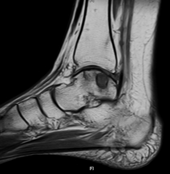
The most challenging aspect of this disease is that it is practically never detected in its initial stages, when it could be managed with less aggressive treatment. This is explained by the fact that routine radiographic examination reveals no changes indicating the presence of necrotic bone tissue within the talus. Consequently, the patient is given a diagnosis that completely fails to address the condition of the talus. The earliest signs of talar necrosis can only be detected via MRI examination, specifically through a detailed and targeted analysis of the talus slices in various imaging sequences. Radiographic and CT signs of talar necrosis only begin to appear when the segments of the necrotic talus undergo lysis (resorption), resulting in the formation of bone cavities or changes in the configuration of the talus due to its destruction.
Two primary factors play a key role in the development of avascular necrosis of the talus: trauma to the ankle joint region and abnormal load distribution on the talus. In the literature, alcohol abuse, prolonged use of hormonal medications, and diabetes mellitus are also cited as contributing factors to the development of avascular necrosis of the talus.
The treatment of talar necrosis is a complex and lengthy process. Commonly used modalities include PRP therapy, shockwave therapy, and magnetotherapy; medications that improve blood circulation are prescribed, and core decompression (tunneling) of the talus or mosaic osteochondral autograft transplantation is performed. Unfortunately, the ultimate outcome of such treatments is typically the same: necrosis of the talus continues to progress, concluding in a debilitating surgery—ankle arthrodesis (joint fusion), which eliminates movement in the joint. In some clinics across Europe and the USA, total ankle arthroplasty was performed to preserve joint mobility, but due to a high rate of complications and failures, this procedure has been practically abandoned.
This naturally raises the question: "Is avascular necrosis of the talus a final sentence, and is disability unavoidable?" No. This is not the case.
To successfully treat avascular necrosis of the talus, three primary objectives must be achieved. First, blood circulation within the necrotic region of the talus must be restored. Second, the necessary conditions for the regeneration of the talar bone tissue must be established. Third, the load distribution on the talus must be normalized. Achieving these three objectives allows for the resolution of avascular necrosis of the talus and the preservation of the ankle joint.
We have developed a technology for staged surgical treatment of avascular necrosis of the talus that addresses all three objectives. This approach integrates three distinct methodologies. To restore blood supply to the talus, we utilize the dorsalis pedis artery (vascular technology). To reconstruct the necrotic segment of the talus, we perform stem cell transplantation or grafting of a segment of the patient's autologous bone (transplantation technology). To normalize the load distribution on the talus, we perform a wedge resection of the tibia (orthopedic technology).
This technology was first applied in 2007 in a patient presenting with near-total necrosis of the talar dome. Fortunately, the structural shape of the talus was completely intact. Six months after the completion of treatment, the bony structure of the talar dome had completely regenerated, and another six months later, the patient was able to utilize his ankle joint normally. In subsequent years, this technology was successfully implemented in 72 patients. In all of these individuals, avascular necrosis of the talus was completely eradicated. None of them required ankle arthrodesis (joint fusion), and only 5 patients retained minor limitations in the range of motion of the ankle joint.
Clinical cases
The girl had Avascular necrosis of the talus was diagnosed. She consulted in various medical institutions, including Germany. They suggested performing an operation to remove the dead section of bone and arthrodesis (closure) of the ankle joint, after which there would be no movement in the joint.
We have implemented restorative surgical treatment using cellular technologies (stem cells).
In six months After the operation, the bone structure of the talus was completely restored. The previously disturbing pain in the joint has completely gone away, and movements in the ankle joint have been restored to full extent.

The woman contacted us for avascular necrosis of the right talus. In 2018, she received a comminuted fracture of the right talus. At the Kiev Institute of Traumatology, the patient underwent osteosynthesis of the fracture. After the operation, the patient continued to have severe pain in the ankle joint. Eight months after surgery, she was diagnosed with avascular necrosis of the talus and was offered ankle arthrodesis. The patient was consulted in clinics in Belgium, Austria, Germany and the USA, and in all clinics she was offered to undergo joint arthrodesis or an artificial joint and were told that she would no longer be able to walk normally.
During examination Total necrosis of the talus trochlea and the absence of cartilage tissue were revealed. The patient was offered a staged surgical treatment developed by us with vascularization of the talus and the use of stem cells. We started treatment almost a year after identifying avascular bone necrosis.
After seven months After transplantation, new bone tissue formed in the area of necrosis. The pain completely disappeared and the patient began to develop movements in the ankle joint and gradually load her leg. Subsequently, we injected stem cells into the joint cavity twice, which created conditions for the formation of cartilage tissue.
Currently The patient walks independently, movements in the joint have been restored almost completely, there is no pain in the joint.

A 24-year-old young woman presented to our clinic with a diagnosis of avascular necrosis of the right talus. This diagnosis was first established 2 years ago. Regarding the necrosis, Alina underwent surgery at a specialized medical institution, where an osteochondral autograft transplantation of the necrotic area of the talus was performed. However, regeneration of the talar tissue did not occur, and the patient was advised to undergo ankle arthrodesis ("fusing" the joint). She declined this surgical procedure because she did not want to be disabled at 24 years of age.
Upon examination, we detected a defect in the medial dome of the right talus, substantial bone marrow edema indicating a significant zone of necrosis within the talus, and an impairment of the talar load distribution.
We performed staged surgical treatment for Alina. During the first stage of surgical treatment, resection of the necrotic segment of the talus was performed, combined with bone autografting and vascularization of this region. In the second stage, correction of the axial load of the talus was carried out.
Seven months after the initiation of treatment, the talus had completely regenerated, and Alina was able to fully utilize her right ankle joint.
Upon examination 2.5 years postoperatively, the talus is completely intact (normal). The range of motion in the ankle joint is full. The patient leads an active life, has married, and is planning a pregnancy.
In June 2019, a 24-year-old patient came to us with complaints of constant aching pain in the ankle joint, which intensified while walking. During examination, she was diagnosed with avascular necrosis of the talus with lysis of the inner upper corner of the trochlea of the talus and valgus alignment deformity of the articular surface of the lower third of the tibia. The defect of the articular surface of the talus trochlea was almost 30%.
Before contacting us the patient was treated at one of the institutes of traumatology in Ukraine. At the beginning of 2015, she underwent mosaic chondroplasty of the area of aseptic necrosis of the talus using osteochondral autografts from the lateral condyle of the femur. Then, after some time, a platelet suspension (PRP therapy) was administered twice into the cavity of the ankle joint. However, pain in the ankle joint persisted and, according to the patient, even intensified. The grafts were completely lysed, and an osteochondral defect formed along the inner part of the articular surface of the talus trochlea.
We have completed vascularization of the area of necrosis of the talus with bone autoplasty from the iliac wing, osteotomy of the tibia with elimination of valgus deformity. Subsequently, to restore articular cartilage, mesenchymal stem cells in the form of a suspension were transplanted into the joint cavity. The transplant was completely rebuilt and formed new bone tissue, over which the stem cells formed new articular cartilage (see figure).
Currently The patient has full use of her leg. According to the patient, pain in the ankle joint does not bother her; sometimes minor pain appears after prolonged exercise (when she has to spend the whole day on her feet) and before a change in weather.

A 61-year-old male patient presented to our clinic with a diagnosis of avascular necrosis of the right talus. This was a repeat consultation. He had previously sought our advice 3 years prior, but had declined the proposed surgical treatment at that time. Throughout these years, he consulted various specialists who administered different modalities of conservative management: intra-articular injections of PRP and hyaluronic acid, shockwave therapy, and other treatments. However, despite the treatment, the pain in his right ankle joint steadily worsened, which significantly restricted the patient's mobility.
Upon objective evaluation, we detected necrosis of the posterolateral dome of the right talus, bone marrow edema, and a load distribution impairment of both the right tibia and the right talus.
The patient underwent staged surgical treatment. During the first stage of the surgical intervention, correction of the mechanical load axis of the tibia and talus was performed. In the second stage, we performed excision of the necrotic segment of the talus combined with bone autografting and vascularization of this region.
One year after the initiation of treatment, the talus had completely regenerated, and Volodymyr experiences no pain whatsoever in his right ankle joint. Volodymyr was able to fully resume an active lifestyle.
